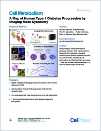
A Map of Human Type 1 Diabetes Progression by Imaging Mass Cytometry.
- Damond N Institute of Molecular Life Sciences, University of Zurich, Zurich, Switzerland.
- Engler S Institute of Molecular Life Sciences, University of Zurich, Zurich, Switzerland.
- Zanotelli VRT Institute of Molecular Life Sciences, University of Zurich, Zurich, Switzerland; Systems Biology PhD Program, Life Science Zurich Graduate School, ETH Zurich and University of Zurich, Zurich, Switzerland.
- Schapiro D Institute of Molecular Life Sciences, University of Zurich, Zurich, Switzerland.
- Wasserfall CH Department of Pathology, Immunology, and Laboratory Medicine, College of Medicine, University of Florida, Gainesville, FL, USA.
- Kusmartseva I Department of Pathology, Immunology, and Laboratory Medicine, College of Medicine, University of Florida, Gainesville, FL, USA.
- Nick HS Department of Neuroscience, College of Medicine, University of Florida, Gainesville, FL, USA.
- Thorel F Department of Genetic Medicine and Development, iGE3 and Centre facultaire du diabète, Faculty of Medicine, University of Geneva, Geneva, Switzerland.
- Herrera PL Department of Genetic Medicine and Development, iGE3 and Centre facultaire du diabète, Faculty of Medicine, University of Geneva, Geneva, Switzerland.
- Atkinson MA Department of Pathology, Immunology, and Laboratory Medicine, College of Medicine, University of Florida, Gainesville, FL, USA.
- Bodenmiller B Institute of Molecular Life Sciences, University of Zurich, Zurich, Switzerland. Electronic address: bernd.bodenmiller@imls.uzh.ch.
- 2019-02-05
Published in:
- Cell metabolism. - 2019
T cell recruitment
endocrine pancreas
highly multiplexed imaging
imaging mass cytometry
immune cells
in situ imaging
islet of Langerhans
pseudotime
type 1 diabetes
β cell
Biomarkers
Diabetes Mellitus, Type 1
Disease Progression
Humans
Image Cytometry
Insulin-Secreting Cells
Islets of Langerhans
Pancreas
English
Type 1 diabetes (T1D) results from the autoimmune destruction of insulin-producing β cells. A comprehensive picture of the changes during T1D development is lacking due to limited sample availability, inability to sample longitudinally, and the paucity of technologies enabling comprehensive tissue profiling. Here, we analyzed 1,581 islets from 12 human donors, including eight with T1D, using imaging mass cytometry (IMC). IMC enabled simultaneous measurement of 35 biomarkers with single-cell and spatial resolution. We performed pseudotime analysis of islets through T1D progression from snapshot data to reconstruct the evolution of β cell loss and insulitis. Our analyses revealed that β cell destruction is preceded by a β cell marker loss and by recruitment of cytotoxic and helper T cells. The approaches described herein demonstrate the value of IMC for improving our understanding of T1D pathogenesis, and our data lay the foundation for hypothesis generation and follow-on experiments.
- Language
-
- English
- Open access status
- bronze
- Identifiers
-
- DOI 10.1016/j.cmet.2018.11.014
- PMID 30713109
- Persistent URL
- https://sonar.ch/global/documents/175719
Statistics
Document views: 28
File downloads:
- fulltext.pdf: 0